AIM:
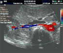
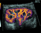
To examine with ultrasound the arteries that branch off the abdominal aorta to supply the kidneys with blood. These arteries can become narrowed (stenosed) or abnormally dilated (aneurysm). Narrowing can cause loss of renal function or high blood pressure while aneurysms can burst and bleed if allowed to enlarge. The ultrasound examination employs sound waves to examine the shape of the artery and to study the flow inside the artery (i.e.; 2 types of information and thus the name ‘Duplex’ ultrasound). This information about an artery helps your doctor to make a diagnosis and decide treatment without using needles or invasive tests. This type of ultrasound can only diagnose and does not treat the arterial problem.
PREPARATION:
The test requires that the subject fasts for 6 to 8 hours beforehand as swallowed food and gas in the gut will obscure the technician’s view of the kidneys, which are deep in the abdomen near the spine.
TECHNIQUE:
The subject lies on an examination couch with just the abdomen exposed. The technician applies ultrasound jelly and presses the ultrasound probe on the abdominal wall, angling it in order to direct the sound beam in various directions. The probe detects reflected sound waves after they bounce off various organs in the belly and images are produced on the computer screen. (see examples below)
DURATION:
Approximately 60 minutes.
DIAGNOSTIC CRITERIA:
A greater than 60% stenosis is predicted if the ratio of the peak velocity in the stenosed renal artery to the velocity in the aorta is greater than 3.5.
Click here to view the worksheet.