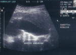
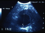
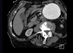
AIMS:
This test is commonly performed for the assessment of exercise-induced leg pain or suspicion of reduced pulses in the lower extremities. It is also done to assess the results of balloon angioplasty or stenting of the iliac arteries and following some iliac artery bypass operations. Ultrasound is used to show location and extent of atheroma (plaque formation), critical stenosis or occlusion, by producing detailed images and blood flow data from the abdominal aorta and its major pelvic branches.
PATIENT PREPARATION:
For morning scan appointments, fast from midnight the night before the examination (no food, fluid, no smoking, no chewing gum). For afternoon appointments, fast for 8 hours prior to your scan time. Diabetics using insulin should not fast. Diabetics not using insulin should notify us when booking the test, and we will endeavour to make your appointment early in the morning. If this is possible, we would like you to fast as above. All patients should take their usual oral medications with a small amount of water.
TECHNIQUE & TEST DURATION:
The test takes about 45 – 60 minutes. The skin over the abdomen and groins is coated with a small amount of clear jelly and an ultrasound probe is pushed against the skin. This produces a picture of the aorta and its branches, and also measures blood flow. Accurate measurements are made of the size of the arteries and blood flow velocities. Sometimes measurements of the pressure and blood flow in the legs is done at the same time as this test using blood pressure cuffs placed around the calf and thigh.
DIAGNOSTIC CRITERIA:
The presence and extent of plaque formation or vessel blockage is assessed by the appearance of the arteries on the ultrasound screen, and also the characteristics and speed of blood flow through the arteries. Generally, as arteries become more blocked, the speed of blood flow increases, and the flow becomes very turbulent and inefficient. Extent of blockage is reported in ranges such as <50%, 50 – 74%, 75 – 99% or completely occluded.
Click here to view the worksheet.


